October 2026 - 1
Author: Dr Joshua Kirk
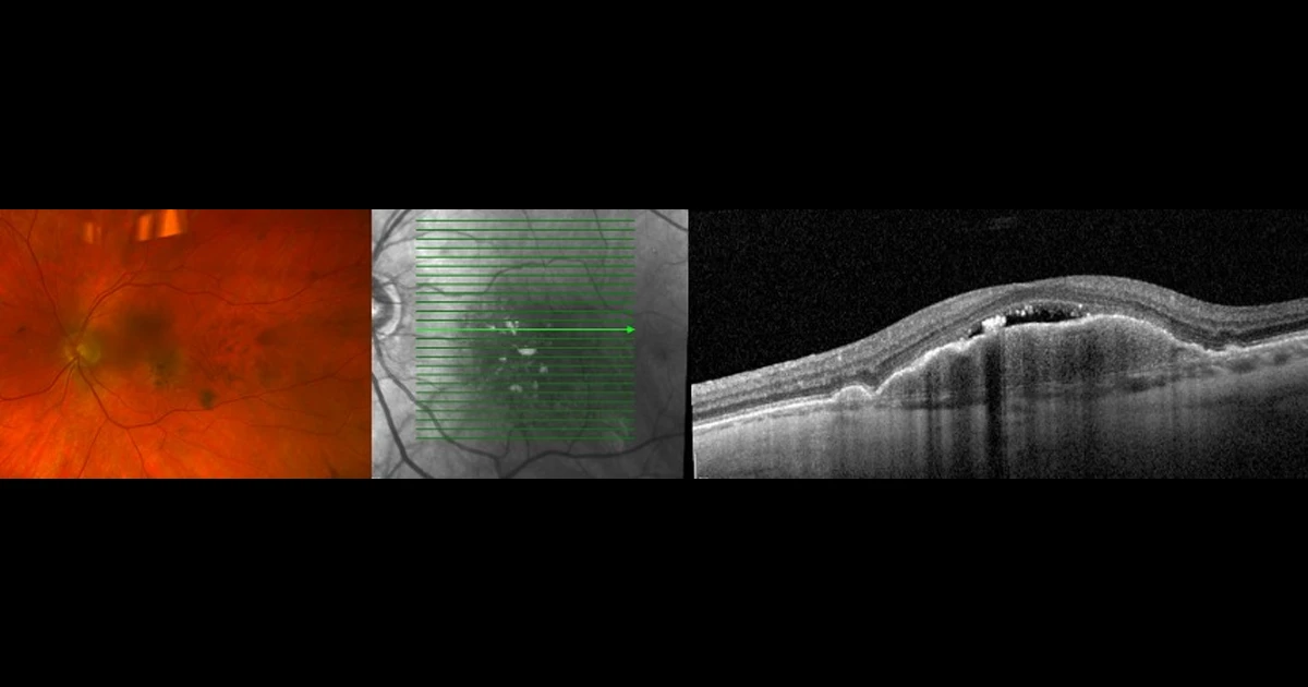
A 65 year old gentleman presented with a 2 week history of mild distortion in his left eye. VA was 6/12 in the affected left eye and 6/9 in the fellow eye. This was on a background of intermediate AMD. Fundus photographs show a large yellowish drusenoid PED with overlying pigment deposition. No haemorrhage is seen. OCT shows a corresponding large PED with overlying subretinal hypo-reflective space.
Differentials include:
A - Neovascular AMD
Oops! Try again
B - Adult Onset Vitelliform Dystrophy
Oops! Try again
C - Non-neovascular AMD with subretinal fluid
Oops! Try again
D - A and C
CORRECT!
Answer D
This particular patient has non-neovascular AMD with subretinal fluid. OCT angiography did not reveal any vascular networks suspicious for a CNV. However, given the patients new visual symptoms, it was decided to trial a course of anti-VEGF injections after a discussion of the risks and benefits. There was no change to the OCT appearance after three injections so a joint decision was made to cease treatment. His vision remained stable at 6/12 and there was no worsening distortion.
Non-neovascular AMD with subretinal fluid is a well described phenomenon (1). The OCT typically demonstrates three different morphologies:
i) Crest of fluid over the apex of a drusenoid PED (as seen in this case),
ii) Pocket of fluid at the angle of a large druse or PED or
iii) Fluid in the depression between confluent drusen or drape of fluid overlying confluent drusen.
It is thought that the presence of subretinal fluid in these settings is due to RPE pump failure. Subretinal fluid in the absence of CNV is an important clinical entity to recognise to prevent the unnecessary use of anti-VEGF therapy.
References
1. Hilely A, Au A, Freund KB, et al. Non-neovascular age-related macular degeneration with subretinal fluid. British Journal of Ophthalmology 2021;105:1415-1420.