
November 2025 - 1

Author: Dr Dov Hersh
An 84 year old female was referred in with decreased left vision LVA 6/18 pH 6/15. Past ocular history bilateral cataract surgery 7 years prior with post op vision RVA 6/7.5 LVA 6/7.5. Systemically well. Whilst taking the history from the patient an area of thinned sclera with underlying pigment was noticed on the left medial conjunctiva (Image above).
Differential diagnoses include?
A - Scleral necrosis post B irradiation for pterygium
Oops! Try again
B - Scleral extension of uveal melanoma
Oops! Try again
C - Post scleritis atrophy
Oops! Try again
D - All of the above
CORRECT!
Answer D
On further examination the patient was found to have a large pigmented choroidal mass taking up most of the superior fundus. B-Scan ultrasound displayed a large choroidal mass with low internal reflectivity a finding highly correlated with choroidal melanoma. Prognosis depends on tumour size, cytogenetic profile, and presence of metastases, with the liver being the most common site of distant spread.
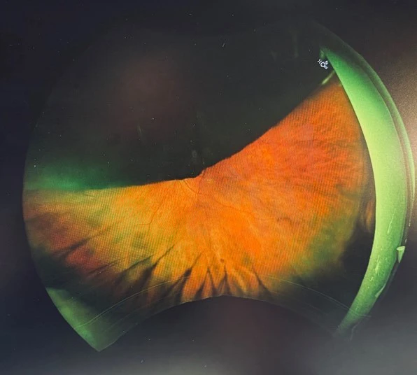
Large superior pigmented choroidal mass
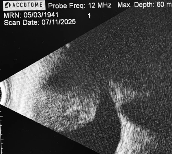
B-Scan ultra sound displaying a large choroidal lesion with low internal reflectivity – consistent with a choroidal melanoma
B irradiation, necrotising scleritis and choroidal melanoma can all present with scleral thinning and underlying pigmentation. A through history and dilated examination including wide field imaging must be carried out to ascertain the aetiology.