
November 2024
Author: Dr Dov Hersh
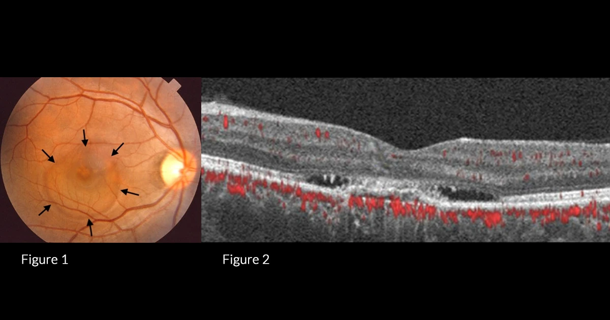
A 62 year old male presented with decreased vision and loss of inferior visual field right eye.
RVA 6/12, LVA 6/5. The fundus images and OCT are displayed below. The fundus image (Figure 1) displays a subretinal blister of fluid (black arrows) with disruption of underlying RPE at the fovea; The OCT (Figure 2) displays subretinal fluid with disruption of the RPE and ellipsoid zone.
Which of the following is not on the list of differential diagnoses?
A - AMD/CNV
Oops! Try again
B - CSR
Oops! Try again
C - Posterior scleritis
Oops! Try again
D - Diabetic macular oedema
CORRECT!
Answer D
The patient presents with sub-retinal fluid and associated RPE changes. The most likely differential diagnoses in this patient demographic are choroidal neovascular membrane (CNV) and central serous retinopathy (CSR). Fundus fluorescein angiogram (FFA, Figure 3) and Fundus Auto Fluorescence (FAF, Figure 4) were performed to help further delineate between CSR and CNV, however were both non-specific.
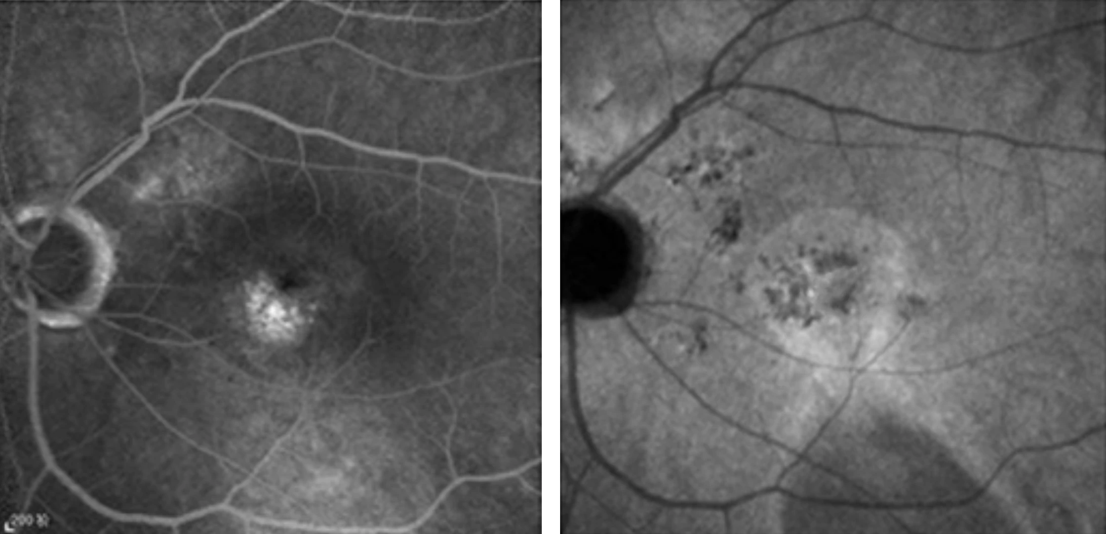
Figure 3
Figure 4
An OCT-A was performed and displayed a clear neovascular membrane in both the superficial (Figure 5) and deep vessel layer (Figure 6), confirming the diagnosis of CNV rather than CSR.
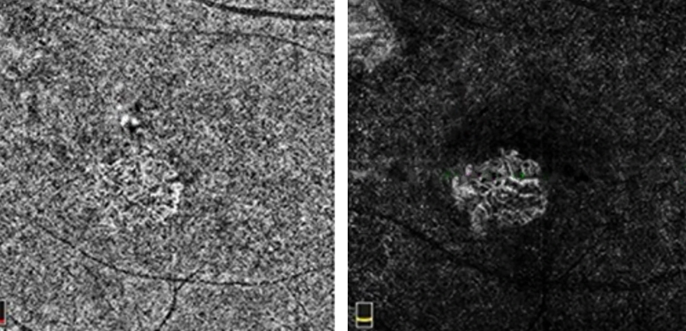
Figure 5
Figure 6
Optical Coherence Tomography Angiography (OCT-A) is a non-invasive, dye-free imaging technique effective for visualising retinal blood vessels and detecting microvascular changes in diseases such as diabetic retinopathy and AMD. As a relatively new technology, OCT-A is proving useful in clinical practice, aiding in the differentiation of specific retinal vascular conditions. Its role in ophthalmology continues to expand as its applications become clearer. However, OCT-A can be sensitive to motion artifacts, which is a limitation to consider.