
May 2025 - 1
Author: Dr Alannah Walsh
Patient A: A 36-year-old Thai woman presented to Sydney Eye Hospital with a six-month history of blurred vision. Her BCVA was R CF and L 6/15. Her IOPs measured at R 58mmHg and L 62mmHg. Her anterior segment was unremarkable. Her fundus examination revealed advanced disc cupping bilaterally. A visual field was unable to be performed in the right eye due to her inability to fixate. The left eye was unreliable.
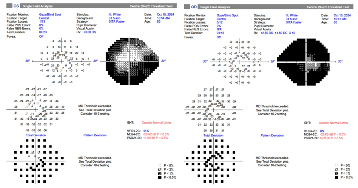
Patient B: An 86-year-old Indonesian man was referred with foggy vision for cataract and glaucoma assessment. His BVCA was R 6/21 and L 6/9. His IOPs measured at R 21mmHg and L 20mmHg. His anterior segment revealed significant cataracts bilaterally but was otherwise unremarkable. Fundus examination revealed advanced disc cupping bilaterally. His visual fields showed advanced constriction (see above).
What is the most valuable next step in these patient’s clinical assessment?
A - Central corneal thickness
Oops! Try again
B - Assessment of the pupillary margin and anterior lens capsule for PXF material
Oops! Try again
C - Gonioscopy
CORRECT!
D - Retro-illumination of the iris for trans-illumination defects
Oops! Try again
Answer C
Following gonioscopy, both these patients were diagnosed with advanced primary angle closure glaucoma (PACG).
Patient A had 360 degrees of synechial closure in both eyes. Her intra-ocular pressures were medically lowered and bilateral peripheral iridotomies were performed. Subsequently, bilateral phaco/trabeculectomies were performed to definitively manage her pressure. Due to the presence of complete synechial angle closure, clear lens extraction alone would have been insufficient to control her IOP.
Patient B had 360 degrees of appositional angle closure. He was commenced on latanoprost to lower his IOP and bilateral cataract surgery was performed. This had the effect of definitively opening his anterior chamber angle and improving his vision. Postoperatively, his IOP measured 9mmHg in both eyes and his VA improved to 6/6 bilaterally.
Patients at increased risk of PACG include those with hyperopia, a family history, advancing age, females, people of Asian or Inuit descent, or those with shallow anterior chamber depths, short axial lengths or increased lens thickness.
PACG is an insidious form of glaucoma. Its effects can be devastating if not identified and addressed appropriately. Early detection and intervention are the key to halting glaucoma progression and preserving vision in patients with PACG.