
September 2024
Author: Dr Alex Hamilton
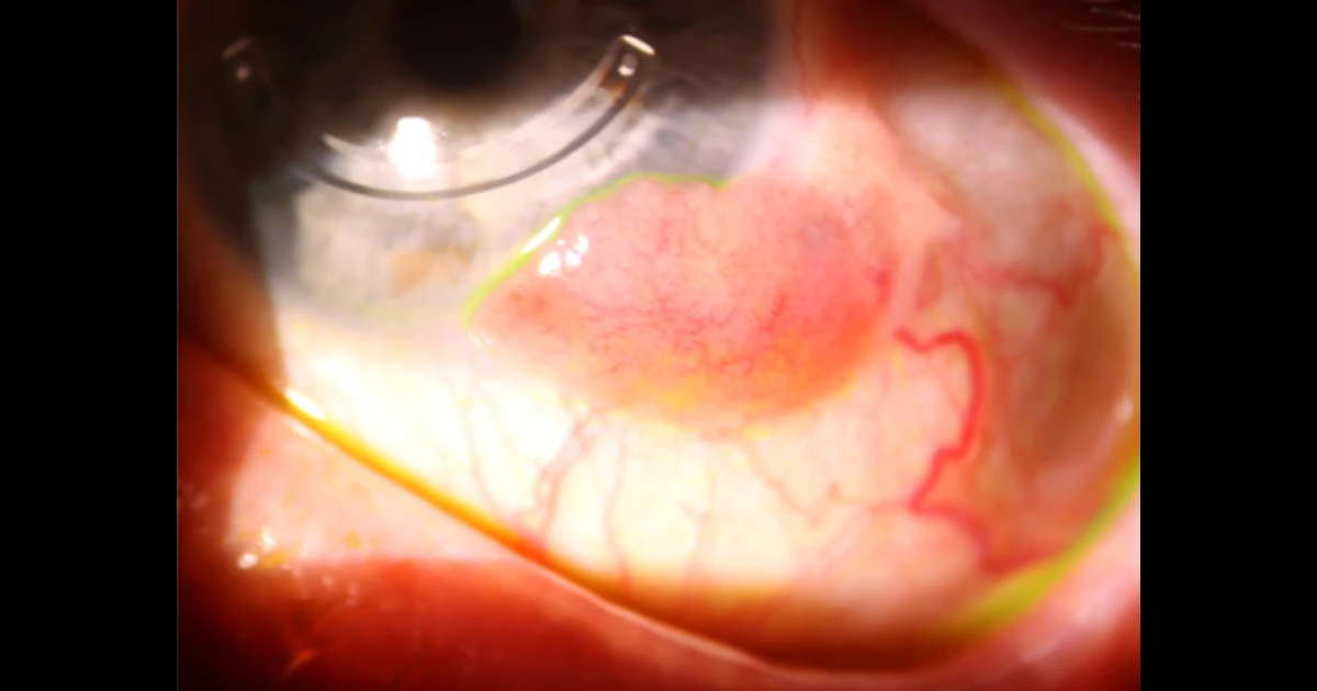
A 62 year old otherwise healthy male presented with a left limbal lesion.
He first noticed the lesion approximately 1 year earlier and noted it gradually increasing in size. He was troubled by mild redness and irritation.
He had a background of keratoconus and has an INTACS corneal implant that was placed many years ago.
Which of the following is not on the list of differential diagnoses?
A - OSSN
Oops! Try again
B - Basal cell carcinoma
CORRECT!
C - Invasive SCC
Oops! Try again
D - Amelanotic melanoma
Oops! Try again
Answer B
A relatively new, large limbal lesion should be referred to an ophthalmologist.
The clinical appearance here was most in keeping with OSSN. As such, surgery was performed to excise the lesion. Double freeze thaw cryotherapy was performed.
The specimen was sent to a laboratory for histopathology. The diagnosis was confirmed as Amelanotic Melanoma.
Amelanotic melanoma is a very rare tumour. This patient requires multidisciplinary care and, in this case, needs a radioactive plaque (Brachytherapy) as further treatment.
This case highlights the importance of a tissue diagnosis to confirm the pathological entity causing a limbal lesion. Only with this tissue diagnosis can appropriate further treatment be undertaken.