
June 2025 - 1
Author: Dr Jeremy Tan
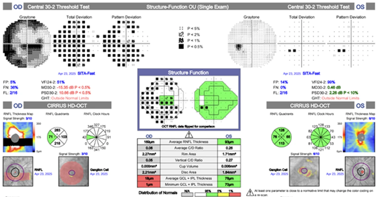
A 62-year-old woman presented with a 5-day history of painless right visual loss.
She denied headache, scalp tenderness, jaw claudication, or systemic symptoms. Her BCVA was R 6/12 and L 6/7.5. Examination revealed a right RAPD and a swollen right optic disc. HVF 30-2 showed a diffuse defect in the right eye. ESR and CRP were normal.
What is the most appropriate next investigation?
A - CT orbit
Oops! Try again
B - MRI brain and orbits with contrast
CORRECT!
C - Temporal artery biopsy
Oops! Try again
D - Lumbar puncture
Oops! Try again
Answer B
This patient presents with unilateral optic disc swelling and visual field loss, raising concern for non-arteritic anterior ischemic optic neuropathy (NAION). However, the differential diagnosis for unilateral disc swelling is broad and includes:
- NAION (most common in older adults with vascular risk factors)
- Optic neuritis
- Compressive optic neuropathy
- Infiltrative or inflammatory causes
- Papillitis or early papilledema (if bilateral)
- Giant cell arteritis (GCA) — a critical diagnosis to exclude in patients over 50
In this case, MRI brain and orbits with contrast was performed and revealed a right paraophthalmic internal carotid artery (ICA) aneurysm adjacent to the optic nerve, raising the possibility of compressive optic neuropathy. This highlights the importance of neuroimaging in all cases of atypical optic neuropathy, especially when:
- no pain (unusual for optic neuritis)
- progressive or severe vision loss
- disc swelling without clear cause
- neurological symptoms or RAPD