June 2024 - 1
Author: Dr Dov Hersh
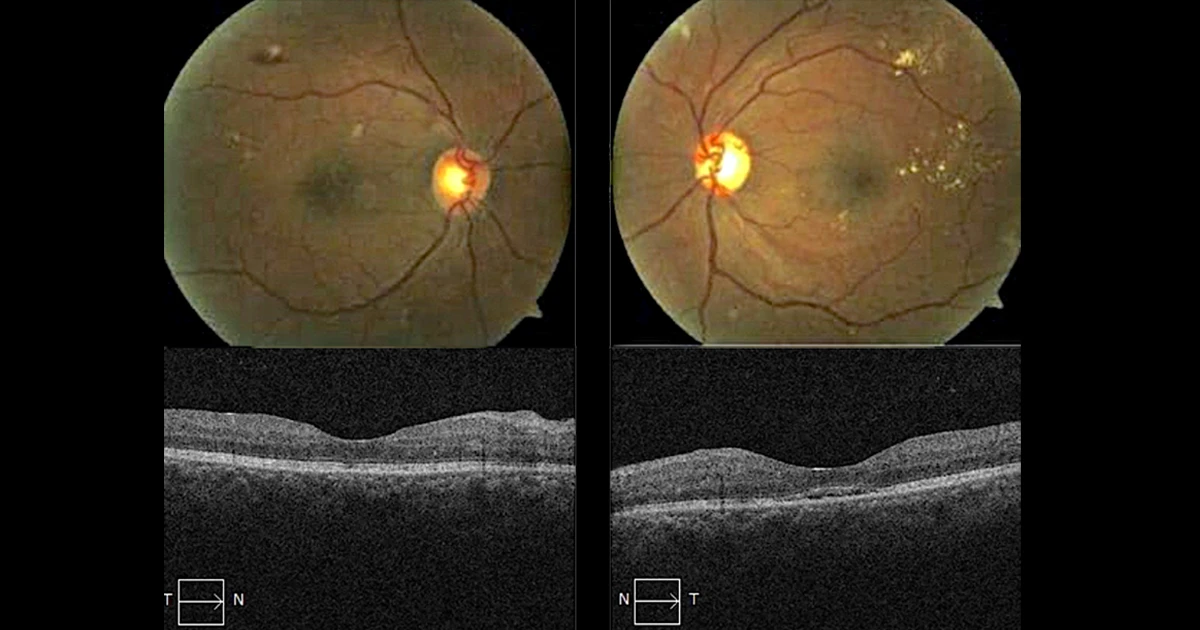
A 23 year old male presented with sudden onset of blurred vision.
He has a 5 year history of well controlled type 2 diabetes with a HbA1c of 5.5. Left VA 6/12, Right VA 6/24. The fundus photos and OCT are displayed above.
What is the next investigation that should be performed?
A - Immediate blood sugar level
Oops! Try again
B - Fluorescein angiogram
Oops! Try again
C - Blood pressure
CORRECT!
D - Temperature
Oops! Try again
Answer C
The patient presents with bilateral retinopathy and macula oedema. Although this clinical picture is often seen in diabetic patients, it is highly unusual for retinopathy and maculopathy to occur in patients with a history of well controlled type 2 diabetes. The rapid onset of decreased vision with the clinical findings above should prompt a differential diagnosis including hypertensive retinopathy. A blood pressure measurement in the clinic or via the patient's GP should be undertaken to exclude systemic hypertension as a cause for the retinopathy. If left systemically untreated, patients with this degree of hypertensive retinopathy are at very high risk of stroke or heart attack. Fluorescein angiography may be warranted at a later time than at the initial presentation to assess for ischaemia and neovascularisation. Although systemic infections such as Bartonella can cause neuroretinitis this is far down the list of differentials and presents somewhat differently. It would be sensible to check the patient's blood sugar levels, but the priority is to exclude a hypertensive crisis.
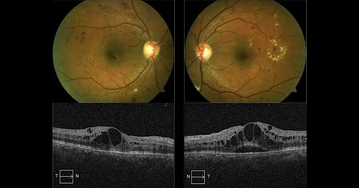
The patient's blood pressure was taken in the clinic and found to be 220/110. The patient was sent to the local hospital emergency room, where he was found to have extremely high blood pressure and was in acute renal failure due to this. His blood pressure was gradually brought down to 120/78 over 8 days. At the 4 week ophthalmic review visual acuity had improved to 6/9 in both eyes and there was a marked improvement in the retinopathy and oedema without direct intraocular intervention.
Post treatment fundus photos and OCT
Use slider to compare with pre treatment images